Endodontic management of maxillary lateral incisor with palato-gingival groove - an innocuous culprit of endo-perio lesion.
Dr. Rafeza Sultana
MS-Resident, Phase-B
Dept. of Conservative Dentistry & Endodontics
BSMMU
Abstract:
Morphological defects occurring in dental structures can be sometime predisposing factor for the onset of inflamatory processes in the periodontal &/ pulpal tissue. Palato-gingival groove is one such defect, most frequently found on the palatal surface of the maxillary laterals. Recognition of such a defect is critical & important, especially because of its diagnostic complexity & further consequences. This case repot is to describe the clinical management of tooth with palato-gingival groove (PGG) in a right maxillary lateral incisor with endo-perio lesion leading to dentoaiveolar abscess & sinus tact.
Keyword: Palato-gingival groove, Endo-perio lesion.
Introduction:
The region where the maxillary lateral incisors are located is considered to be an area of embryological hazard (J Dijalma et al, 1991). A great number of minor & major malformations occur in this area. For instance cleft palate, globule-maxillary cyst, missing or supernumerary tooth, dense in dente & peg lateral incisors. Another anomaly occurring in that region is PGG (Everetti FG et al, 1972).
The etiology of groove formation is not fully understood. But it is thought that the formation of PGG is by infolding of the enamel organ & the Hertwing’s root sheath (Walker & Jons, 1983) & has been speculated to an aborted formation of additional root (Keruzoudis et al, 2003).
The PPG has the similarity to dens invaginatus, however it differs from dens invaginatus in such way that PGG occurs due to an infolding of the epithelium (resulting in a groove), rather than an invagination (resulting in a circular opening) (TG Ground 1988).
The anomaly has a variety of name, the PPG, the radicular lingual groove, the palato-radicular groove, the facial-radicular groove, the disto-lingual groove (Cacflia MS et al, 1998).
The PPG is the funnel shaped appearance which forms a niche for bacterial plaque & calculus accumulation making it difficult for the patients as well as professionals.
These grooves can be classified in to mild, moderate & complex based on its depth and extent. Mild one terminates at the CEJ whereas moderate grooves continue apically along the root surface. The complex forms are deeply grooved defects that separate an accessory root from the main root trunk (Goon WW et al, 1991).


Mild Moderate Complex
Fig: Different types of palato-gingival groove.
Deferent studies have revealed a prevalence rate for palatal groove of about 2.8% to 8.5%, the most prevalent being the maxillary lateral incisor (D Rachana et al, 2007). In lateral incisors 43% of the grooves on the root extended less than 5mm. 47% between 6-10mm and only 10% more than 10mm (Kogan 1986).
Case report:
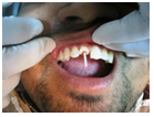
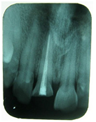
A 30 year old female patient came to the dept. of conservative Dentistry & Endodontics, BSMMU with the complaint of occasional episodes of swelling of gum & purulent discharge in relation to upper right lateral incisor. Her medical history was non contributory. There was no history of trauma, caries nor any discoloration of tooth. Intra-oral examination showed localized swelling & an intraoral draining sinus pointing on the labial gingiva at the apex of right lateral incisor. The palatal surface of lateral incisor showed fossa with mild calculus embedded in it. Periodontal examination revealed mild bleeding on probing & a narrow periodontal pocket (<5mm) alongside the groove & other aspects were revealed normal. There was no mobility associated with it. To locate the origin of the sinus, a gutta percha cone (no#25) was inserted in to its course & a radiograph was taken. The GP cone pointed towards the apex of the offending tooth. Radiograph also showed periapical radiolucency (about 3mm). On vitality test the tooth was found to be non vital. The findings were suggestive of primary pulpal lesion (Type-1 endo- perio lesion).
So, the case was diagnosed as type-I endo-perio lesion due to palato-gingival groove (Mild type).
Treatment plan comprised with of oral prophylaxis followed by primary endodontic management provided three dimensional obturation & repair of PGG with glass ionomer cement (Type-II).
The consent of the patient was taken. After mouth preparation, a straight line access cavity was done. The necrotic pulp remnants were removed & washed out by 2.5% NaOcl & normal saline. Working length (WL) measuring radiograph was taken & the WL was established as 20mm. Then the canal was prepared up to no #50 K file along with copious irrigation with NaOcl & normal saline. The canal was dried with paper point & Ca(0H)2 paste was placed as an intracanal medicament for one week. The access was tightly sealed with ZOE cement. At the next visit the canal was found dry. Then the canal was irrigated with normal saline and dried with paper point. Before obturation a master point was inserted in to the canal & felt the tug back. Then the tooth was obturated with ZOE sealer & GP cone by lateral condensation technique and final restoration was made with GIC. The post-operative radiograph was taken at the same visit for obturation evaluation. The PPG was repaired with GIC as well. The patient was advised for follow up at 3, 6 & 12 months intervals.
 |
Fig: exploration of sinus tract
|
 |
Fig: Detection of PGG
|
 |
Fig: Shallow periodontal pocket alongside the grioove
|
 |
Fig: Pre-operative x-ray
|
 |
Fig: Sinus trakcking x-ray
|
 |
Fig: WL-measurring x-ray
|
 |
Fig: Obturation x-ray
|
Discussion:
Palato-gingival groove is a developmental anomaly that has the predilection for the maxillary lateral incisor. The PGG presents of variable extent & depth that may or may not involve a communication between pulp cavity & periodontal tissue. These grooves are deep initially after root formation & become shallow with age due to deposition of dentin (Anderregg CR, 1993). The PPG is one of the important entities & could manifest either as true endodontic lesion, periodontal disease or may appear as a combined endo-perio lesion.
For anatomical region, the PGG is an ideal plaque trap for promoting periodontal break down & pulp necrosis. Reasons for the occurrence of combined lesion are existence of communication between the pulp chamber and the periodontium. Friedman & Goultschin have suggested pulpal necrosis followed by apical periodontitis is often earliest manifestation of PPG.
Clinician should be aware of the incidence & method for treating PPG. Rrarely the PGG can be seen on radiographic examination in the form of a parallel radiolucent vertical line (RW Lee 1968) or in other cases not following the root canal (DS August 1978).
The PGG requires early diagnosis and treatment as it may result in radicular & pulpual pathosis. This fissure like channel is a locus of plaque & calculus accumulation, which acts as a secondary local etiologic factor encouraging the development of periodontitis (Kozlovsky A 1988). A patient with PGG may have the symptoms of a periodontal or acute dento-alveolar abscess or may show no symptoms at all. Frequently a lesion related to a groove is characterized by recurrent symptomatic episodes (Robinson SF 1988).
The pulp is also affected by bacteria which are situated in the radicular groove. Bacteria and their products may enter in to the pulp through the accessory foramen and lateral canals situated along the floor or side walls of the groove. Another groove of the bacterial invasion into the pulp is via the exposed dentinal tubules on the side of the groove where surface resorption as a result of inflamatory process (N P Kerezoudis 2003).
Treatment may vary from case to case. Early diagnosis of the case is very much important for preventive measure. Teeth with deep palatal groove should be treated with fissure sealant before plaque and food impaction & breakdown of the periodontal structures (M Hulsmann 1997).
Although several modalities have been suggested for the treatment of this condition. There is general consensus that these are predictable failures. Many treatment regimen have been suggested such as conventional root canal treatment, combined root canal therapy followed by saucerization of the defect with flowable composite, intentional replantation and guided tissue regeneration according to severity.
In the last decades, with extensive knowledge of guided tissue regeneration, mechanical barrier have been used to halt the epithelium down growth along the root surface, allowing periodontal ligament, cementum and bone to regenerate along periodontally diseased roots. Calcium sulphate, collagen methyl cellulose acetate, enamel matrix protein etc have used as mechanical barrier to allow periodontal regeneration (S Andreana 1998).
The prognosis of a tooth with PPG mainly depends on location of the groove, severity of the periodontal problem, accessibility of the defect & the type of groove ie shallow or deep, long or short.
In this case report, patient presented with true endodontic involvement (type-1 endo-perio lesion), as an earlier manifestation of PGG. So non surgical endodontic treatment was done provided three dimensional obturation & the groove was repaired with GIC & advised for periodic follow up.
GI cement was used because of its antibacterial activity & the property of chemical adhesion to the tooth structure. Clinical & histological studies have been shown that there is an apical & CT adherence to the GI cement during the healing process.
Conclution:
Palato-gingival groove is an enigma & it is considered as a silent killer that can pose dilemmas for diagnosis & clinical management. The PGG might escape detection until patient presents with advanced pulpal pathosis with secondary periodontal involvement. So the evaluation of clinical signs & appropriate diagnostic tests are of paramount important to prevent incorrect diagnosis & treatment. Endodontists must be capable of performing advanced periodontal regeneration techniques during endo-surgery for the successful treatment of those lesions.
References:
Anderegg CR, Meitzer DG (1993) Treatment of the palato- gingival groove with guided tissue regeneration. Report of 10 cases. J Periodontol 64:72-4
Andreana S A (1998) Combined approach of treatment for developmental groove associated periodontal defect. A case report J periodontal 69: 601-7
August DS (1978) The radicular ligual groove: an overlooked differential diagnosis J Am Dent Assoc 90: 1037-9
Cecflia MS, Lara VS, Moraes IG, Paolo S, Brazil (1998) The palato-gingival groove a cause of failure in root canal treatment. Oral Surg Oral Med Oral pathol Oral radiol Endod 85:94-8).
D. Rachana, Prasannalatha Nadig, Gururaj Nadig (2007) The palata groove: Application of computed tomography in its detection – A case report. Journal of conservative dentistry 10: 83-43(6): 353-361.
Everatt FG, Kermer GM (1972) The disto-lingual groove in the maxillary lateral incisor. J Endod 1991: 17:244-8.
Goon WW, Carpenter WM, Braces NM, Ahlfeld RJ, Complex facial radicular groove in a maxillary lateral incisor. J Endod 1991; 17:244-8.
Jesus Dijalma Pecora, Manoel Damia O Sousaneto Et AI (1991) Invitro Study Of redicular Grooves In Maxillary Incisors. Braz Dent J 2(1):69-73.
Kogan S (1986) The prevalence, location and conformation of palato radicular grooves in maxillary incisors. J Peridontol 57:231.
Kozlovsky A, Tal H, Yechezkiely N, mozes O (1988) Facial radicular grooves in a maxillary central incisor – A case report. J Periodontol September 59(9):615-617.
Lee RW, Lee EC, Poon RY (1968) Palato gingival grooves in maxillary incisors: a possible predisposing factoto localized periodontal disease. Brit dent J 2:14-8
M hulsmann (1997) Dens invaginatus: aetiology, classification, prevalence, diagnosis and treatment considerations. Int Endod J 79-90.
N.P. Kerezoudis, G.J. Siskos & V. Tsatsas (2003) Bilateral buccal radicular groove in maxillary incisors: case report. Int Endo Jour 36:898-906.
Robinson SF, Cooley RI (1988) Palato-gingival groove lesions: recognition and treatment. Gen Dent 36:340-342.
Tom G. Gound, Glenn I. Mze (1998) treatment options For The Redicular Lingual Groove A Review & Discussion. Pract Periodont Aesthetic Dent 10(3):369-375.
Reattachment of fractured coronal tooth fragment of maxillary central incisor by using fiber reinforced post- Regaining back to normal.
Dr. Rafeza Sultana
MS-
Resident, phase-B
Dept. of
Conservative Dentistry & Endodontics
Bangabandhu
Sheikh Mujib Medical University, Dhaka.
Abstract:
Dental
trauma is such a situation where in the patient is affected both socially and
psychologically. Such patients are quite apprehensive because of impaired
function, esthetics and phonetics. The prime objective while handling such
cases is immediate restoration of function, esthetics and phonetics as well.
The advances in adhesive dentistry have allowed dentist to use the patient’s
own fragment to restore the fractured tooth. Reattachment of tooth fragment is
such an ultraconservative technique which provides safe, fast &
esthetically pleasing results. This case report presents a 27 year male patient
with a complicated crown fracture of maxillary right central incisor tooth;
where fracture fragment luted with reattachment technique. The procedure used
to repair the fracture fragment included endodontic treatment & after root
canal obturation a glass fiber post is used for reinforcement and fragment was
luted with composite resin. On word assessment showed a stable reattachment,
good esthetic and periodontal health.
Key word: Coronal fracture, fragment reattachment,
Composite resin, fiber post.
Introduction:
Almost
every dental expert is familiar with the patient having traumatized tooth at
their regular practice. The most affected teeth are maxillary incisors due to
their anterior position and protrusion and the common etiological factor of
crown or crown root fracture in the permanent dentition are injuries caused by
fall, contact sports, automobile accident and foreign body striking the teeth.
Esthetic rehabilitation of crown fractures of the maxillary anterior teeth is
one of the greatest challenges to the dental specialist. The patients are very
conscious about their appearance where as the specialist has to consider long
term biological function of that tooth in addition to esthetic. Traditionally
such injuries have been restored with composite resin1, but they have some disadvantages of
colour match and variable wear.6
On the other hand reattachment of fractured fragment may offer following
advantages (1) Better esthetic and achievement of lifelike translucency (2)
Require less time (3) A positive emotional and social response from the
patient’s side5 (4) Relatively
inexpensive procedure.
Moreover
several factors influence the management of coronal tooth fracture including
extend of fracture, pattern of fracture and restorability of fractured tooth,
secondary trauma, presence/absence of fractured tooth fragment and it’s
condition for use, occlusion, esthetic, finances and prognosis.3,15,18 With the recent improvement in the
dental materials, resin based restorative materials with the use of tooth
coloured fiber reinforced polymer posts are of choice for such treatment
protocol. Because the biomechanical properties of fiber reinforced polymer post
are reported close to those of dentin8
like esthetic, bonding to tooth structure, modulus of elasticity and cause
fewer tooth fracture.20
If
a broken tooth fragment is available and in a good condition the restoration of
the tooth using its won fragment has been suggested.21
Case Report:
A
27 year old male patient reported to the Dept. of Conservative Dentistry and
Endodontics BSMMU with the chief complaint of broken upper front tooth
following trauma due to hard substance striking while taking food 7 days back.
His
medical history was all right. Clinical examination revealed a clean fracture
horizontally mesial to distal and angulated incisally from palatal to labial
with pulp exposure on the labial surface of right maxillary central incisor.
Fracture was not evident labially. There no apparent trauma to the adjacent
teeth and soft tissues. On radiographic examination revealed an oblique
fracture palato-labially. After routine history taking, examination and based
upon patient’s desire, a treatment plan was formulated that included endodontic
treatment and reattachment of fractured portion of tooth with composite resin
using a fiber reinforced post.
In
the first appointment a single visit endodontic treatment was performed. Under
local anesthesia, the pulp was extirpated and the working length was determined
by working length measuring X-ray. Then the root canal was prepared as standardized
technique at 17mm working length up to 70 H file and obturated with Gutta
Percha by lateral condensation technique.
The
fractured fragment was completely separated, dehydrated and chalky white in
appearance. In order to prevent dehydration and to get the natural appearance,
the fractured fragment was preserved in normal saline for 7 days. After 7 days,
in the next visit the GP was partially removed by pesos-reamer (No-1) leaving 5
mm GP at the apex to maintain a tight apical seal. A post hole was prepared
within the canal and a perfect diameter sized glass fiber composit
post(Glassix, Nordin) was cemented with the root canal using glass ionomer luting
cement (GC corporation). An internal groove was made both in fractured fragment
and the palatal aspect of the tooth where the fiber post and composite will
occupy. Acid etching was done on both the fragment and the tooth using 37%
orthofhosforic acid for 15 seconds and thoroughly rinse off. A bonding agent
(Beautibond, sofu) was applied to both the substrates and cured according to
manufacturer instruction.
Then the fragment was reattached with flowable
composite resin (Beautifil flow, Sofu). The excess resin was removed with an
excavator and light cured for 30 seconds from both buccal and palatal aspect.
Final finishing and polishing was done. Occlusion was cheeked and post
operating instructions were given and patient was recalled after 7 days for
evaluation. Clinical and radiological examination carried out after 1 month, 3
months, 6 months and 1 year to confirm the satisfactory esthetic and functional
outcome of the treatment with no associated endodontic or periodontal problem.
Discussion:
Up
to date, a lot of deferent approaches were proposed for treatment of fractured
tooth depending on location of the fracture such as (1) Reattachment of the
fractured fragment (2) Composite restoration (3) Orthodontic extrusion (4)
Surgical extrusion (5) Crown lengthening.11
In recent years due to
remarkable advancements of adhesive systems and resin composites, it is now
possible to achieve excellent results with reattachment of tooth fragments
provided that the biological factors, materials, and techniques are logically
assessed and managed.16 As with the conventional restoration, restorative success depends
on proper case selection, strict adherence to sound principles of periodontal
and endodontic therapies, and the techniques and materials for modern adhesive
dentistry.10,12,14
In the presented case of
complicated crown fracture requiring endodontic therapy, the fractured fragment
was available and reattachment of the fragment with fiber post is performed to
retain the fractured segment and reduce the stress on the luting material. The posts
interlock the two separate fragments and minimize the stress on the remaining
tooth structure. The use of the natural tooth substance offers a conservative,
esthetic, and economical option that provides good and long lasting esthetics,
restores function, results in a positive psychological response, and is
certainly a simple procedure. Adhesive post is used as it has the potential for
increased retention, is more flexible, and has modulus of elasticity
approximately same as dentin, and when bonded with resin cement it distributes
forces evenly along the root.17
The most common
complication of post and core system is debonding;4 another reason for
failure is root fracture.9 Restoration with cast metal posts can cause wedging forces
coronally that may result in irreversible failure because of fracture of an
already weakened root.2 Whereas fiber-reinforced composite resin post has demonstrated
negligible root fracture. Studies have indicated that dentin-bonded resin
post-core restorations provide significantly resistance to fracture than
cemented custom cast posts and cores.7,19 In addition; the
fiber-reinforced posts are used with minimal preparation because it uses the
undercuts and surface irregularities to increase the surface area for bonding,
thus reducing the possibility of tooth fracture during function or traumatic injury.13
Various studies reported
that sectional obturation of root canal (at the apex) and use of dual cure
resin play an important role in the successful treatment outcome of
reattachment technique. Use of a fiber post luted with dual cure resin increase
the retention of the segment and provides a monoblock effect by locking the
core material (fiber post) with the dentinal wall of root canal without leaving
any gaps.
Most concerns about
reattachment technique have been directed towards the fractured strength of the
restored tooth. There are several reinforcement techniques adapted to
strengthen the tooth structures like – i) Circumferential bevel, ii) External
chamfer, iii) ‘V’ shaped bevel, iv) Placement of internal grooves, v)
superficial over contour of restorative material over the fracture line and
pulp chamber, in case of complicated fracture.18
The clinician must
consider that a dry and clean working field and proper use of bonding protocols
and bonding materials is the key to achieve success in adhesive dentistry.
Conclusion:
Because of larger
incidence of trauma to dental tissues and their supporting structures, it is
important to have proper knowledge of the techniques available and their
indications, along with risk benefit ratio. The reattachment of the tooth
fragment is possible only when the fragment is available and can be improved
with different adhesive techniques and restorative materials. The main concern
is to educate the population to preserve the fractured fragment and seek immediate
dental care.
References:
1.
Attila IO, Cenk MHA, Serdar MT.
Multidisciplinary approach to the rehabilitation of a crown –root fracture for
immediate esthetics. Dent Traumatol 2006; 22:48-52.
2.
A. S. Deutsch, J.
Cavallari, B. L. Musikant, L. Silverstein, J. Lepley, and G. Petroni, “Root
fracture and the design of prefabricated posts,” The Journal of Prosthetic
Dentistry, vol. 53, no. 5, pp. 637–640, 1985.
3.
Andreasen FM, Noren JG, Andreasen JO, et al.
long term survival of fragment bonding in the treatment of fractured crowns.
Quintessence Int 1995; 26:669-81.
4.
A. Torbjörner, S.
Karlsson, O. Dr, and P. A. Ödman, “Survival rate and failure characteristics
for two post designs,” The Journal of Prosthetic Dentistry, vol. 73, no.
5, pp. 439–444, 1995.
5.
Baratieri LN, Monteiro S. tooth fracture
reattachment: Case reports. Quint Int 1990; 21:261-270.
6.
Badami A, Dunnes, Scheer B. As in vitro
investigation into shear bond strengths of two dentine bonding agents used in
the reattachment of incise edge fragments. Endo Dent Traumat 1995; 11:29-135.
7.
B. Akkayan and T.
Gülmez, “Resistance to fracture of endodontically treated teeth restored with
different post systems,” The Journal of Prosthetic Dentistry, vol. 87, no.
4, pp. 431–437, 2002.
8.
Duret B, Duret F,
Reynaud M. Long-life physical property preservation and postendodontic
rehabilitation with the composipost. Compend Contin Educ Dent
Suppl. 1996;20:50–56.
9.
E. Asmussen, A.
Peutzfeldt, and T. Heitmann, “Stiffness, elastic limit, and strength of newer
types of endodontic posts,” Journal of Dentistry, vol. 27, no. 4, pp.
275–278, 1999.
10.
F. M. Andreasen,
U. Steinhardt, M. Bille, and E. C. Musksgaard, “Bonding of enamel-dentin crown
fragments after crown fracture. An experimental study using bonding
agents,” Endodontics & Dental Traumatology, vol. 9, no. 3, pp.
111–114, 1993.
11.
Georgia. V. Macedo, Patrica Diaz, Carlos
augusto. Reattachment of anterior tooth fragment: A conservative approach.
Journal of Esthetic and Restorative dentistry 2008; 20:5-20.
12.
G. Cavalleri and
N. Zerman, “Traumatic crown fractures in permanent incisors with immature
roots: a follow-up study,” Endodontics & Dental Traumatology, vol. 11,
no. 6, pp. 294–296, 1995.
13.
K. C. Trabert, A.
A. Caputo, and M. Abou-Rass, “Tooth fracture—a comparison of endodontic and
restorative treatments,” Journal of Endodontics, vol. 4, no. 11, pp.
341–345, 1978.
14.
M. N. Lowey,
“Reattachment of a fractured central incisor tooth fragment,” British
Dental Journal, vol. 170, no. 8, article 285, 1991.
15.
Olsburgh S, Jacoby T, Krejci I. crown
fractures in the permanent dentition: pulpal and restorative consideration.
Dent traumatol 2002; 18(3): 103-15.
16.
P. Vashisth, M.
Mittal, and A. P. Singh, “Immediate reattachment of fractured tooth segment: a
biological approach,” Indian Journal of Dental Research and Review, pp.
72–74, 2012.
17.
P. Lokesh and M.
Kala, “Management of mild-root fracture using MTA and fiber post to reinforce
crown—a case report,” Indian Journal of Dental Research and Review, vol.
3, pp. 32–36, 2008.
18.
Reis A, Francci C, Loguercio AD, et al.
Re-attachment of anterior fractured teeth: fracture strength using different
techniques. Oper Dent 2001; 26(3):287-94.
19.
R. T. Beg, M. W.
Parker, J. T. Judkins, and G. B. Pelleu, “Effect of dentinal bonded resin
post-core preparations on resistance to vertical root fracture,” The
Journal of Prosthetic Dentistry, vol. 67, no. 6, pp. 768–772, 1992.
20.
Salameh Z,
Sorrentino R, Papacchini F, Ounsi HF, Tashkandi E, Goracci C, Ferrari M.
Fracture resistance and failure patterns of endodontically treated mandibular
molars restored using resin composite with or without translucent glass
fiber-post. J Endod. 2006;32:7752–7755.
21.
Yilmaz Y, Zehir C, Eyuboglu O, Belduz N.
evaluation of success in the reattachment of control fractures, Dent Traumatol
2008; 24:151-8.
Figures
of various steps of reattachment technique.
 |
| Fig: Pre-Operative photograph |
.png) |
| Fig: Fracture fragment (labial view) |
.png) |
| Fig: Fracture fragment (palatal view) |
 |
| Fig: Fiber reinforced post |
 |
| Fig: post placement |
 |
| Fig: post placement |
 |
| Fig: Groove preparation |
 |
| Fig: Trial of fragment reattachment |
 |
| Fig: Trial of fragment reattachment |
 |
| Fig: Check occlusion |
 |
| Fig: Light curing of composite resin |
 |
| Fig: Light curing of composite resin |
 |
| Fig: Post operative occlusion check |
 |
| Fig: After treatment |
Radiographic
assessment.
 |
| Fig: Pre-operative |
 |
| Fig: per-operative |
 |
| Fig: per-operative |
 |
| Fig: post-operative |
Fluoride seal to forbid dental cavity in children and teenagers
Objective
High dental cavity commons disease affecting a big part of the world wide population, including around 65% to 80% of school-age children. In general, levels of dental cavity varies substantially between and breast of the several countries, but in low socio-economic position (SES), children have higher levels of dental cavity than those in higher SES groups. Treatment of cavity causes of the teeth accompanied by suffering and pain. Repair and alternate of dental cavity is costly, that represents a significant drain on resources for health care systems.
Fluoride seal is a potential option to prevent dental cavity in children. This developed in the 1960s and has been widely used in Europe and Canada. Use in other countries appears to be increasing, including in the US, where it can be used outside of the label as preventive agents of decay.
Two large meta-analysis reviews indicated significant reductions in caries in deciduous and permanent teeth. However, there was significant heterogeneity among the studies within each body of meta-analysis of the evidence for the two meta-analyses of average quality.
Thirteen tests allowed data for meta-analysis of the surface of the permanent tooth, and the fraction bundle of FS (M) prevented estimate comparing fluoride seal with placebo or no treatment was 43% (95% confidence interval (CI) 30-57%). d (e/m) to prevent the perceptiveness of the falling out was 36% (90% CI(: 25% to 52%) Ten tests provided data for meta-analysis of the surface of the tooth. Prevents any significant correlation between the d (M) FS or FS fractions d (e/m) has been found on the baseline of caries risk of infection factors. It was background exposure to fluoride, the capacities of the application such as prevention, fluoride concentration, and continually of application. It there was also no significant association in models meta-regression between estimates of d (M) FS or FS d (e/m) prevents fractures and after special factors such as whether the use of a placebo or no treatment control, duration of follow-up, or whether to use individual or randomized cluster. There was little information on potential adverse effects or accept treatment.
This updated review conclusions remain the same when you appear first, but are enhanced by the addition of various more recent studies. The analysis suggests significant effect prevents caries of primary and permanent dental varnish fluoride, but the quality of the evidence was considered to be moderate, because they included mainly studies present a high risk of bias and heterogeneity.
Endodontic management of Mandibular first molar with five canals
Dr.
Rafeza Sultana
Ms- Resident,
Department of Conservative Dentistry & Endodontic.
BSMMU
Abstract: The purpose of this study was to
demonstrate the importance of knowledge of the internal anatomy of root canals
for the success of endodontic treatment. Lack of knowledge of anatomic
variations & their characteristics in different teeth has been pointed out
as one of the causes of endodontic therapy failure. The present report describes a right mandibular first permanent
molar requiring root canal therapy, found to have three separate canals (
type-1 configuration) in the mesial root
& two separate canals ( type-1 configuration) in the distal root . So
emphasizes the need for the clinicians to be aware of & look out for such
variation & use adequate diagnostic methodologies prior to & during
therapy to detect such variations. The operator experience has also shown to be
a key factor in negotiation & management of this aberrant canal
configuration.
Key Word:
Mandibular 1st
Molar, Middle Mesial canal, % root canal, Root canal anatomy.
Introduction: Knowledge of internal
dental anatomy is fundamental to the success of endodontic treatment. Incomplete
instrumentation, inadequate cleaning & shaping, & the subsequent
defective obscuration of root canals are the main causes of endodontic
treatment failure1. Anatomical characteristics of the different
types of teeth and their possible variations are challenges routinely faced by
practitioners performing endodontic treatment.
The correct access into the pulp
chamber, which should allow access to the orifices of the root canals and an
optimum view of the chamber floor, is a fundamental step in endodontic therapy
as it enables the identification of any variation in the number and position of
root canals2 .
The middle mesial canal has been more
commonly located in mandibular 1st molar10. Several studies have evaluated the degree of
variation in the number of roots and root canals in mandibular 1st
molars10 . Fabra campons10 studies 145 mandibular 1st
molars & found that 2.75% of the teeth had five canals. Martinez-burna and
Badanelli5 conducted a canal investigation & found 29 teeth with
five root canals in a
sample of 2362 mandibular permanent molars and reported that 12 out of 100
molars studied had a third mesial canal.
Aminsobhani et all studied the
occurrence and location of the middle mesial canal of mandibular 1st
molar & second molar in relation to other two mesial canals that were
treated in private practice that middle mesial canalwas located in the middle
of the distance between the mesiobuccal and mesiolingual canals. The canal
configuration was found in 6 2nd
molars & 21 1st molars. Middle mesial canals in all of the cases
joined to mesiobuccal or mesioligualcanals. None of the teeth consisted of three independent canals with three apical
foramina . Beatty & krell described a
mandibular 1st molar with three independent canals in the mesial
root . Author Dr. carlos Heibom et al
reported the number of roots, total number of canals, the number of middle
mesial canals & number of foramina in the following chart
Number of roots
Number of molars studied 18,781 3-rooted molars in
% 13 % (2,450)
|
Total number of
canals
Number of molars studied 4,745 61.3 % 3 canals 35.7 % 4 canals 0.8 % 5 canals
|
Number of canals
in mesial root
Number of mesial roots studied 4,535 3.3 % 1 canal 94.2 % 2 canals 2.6 % 3 canals
|
Mesial and
distal roots. Canal system configuration
Type I (1-1) Type II
(2-1) Type IV (2-2) Type VIII (3-3)
Number of mesial roots studied 4,331 35 % 52.3 % 0.9 %
Number of distal roots studied 2,992 62.7 % 14.5 % 12.4 %
|
Number of
foramina in mesial and distal roots
1 foramen 2
foramina 3 foramina
Number of mesial roots studied 4,817
38.2 % 59.2
% 1.6 %
Number of distal roots studied 3,378 77.2 % 22.2 %
|
Intercanal
communications. Type V isthmuses
Mesial root Distal root
Number of molars studied 1,615 54.8 % middle & apical
1/3 20.2 % middle 1/3
|
According to ingle12 one
of the most important causes of endodontic treatment failure is the incomplete
obturation of the root canal system. Therefore, the correct location,
instrumentation & obturation of all canals are indispensable
procedures.
Case report: A 19 years old male patient reported
to the department of conservative dentistry & endodontics BSMMU with
decayed tooth & associated pain over his right mandibular region. Intra
oral examination revealed class 1 deep carious lesion in mandibular right 1st
molar. The tooth exhibited no mobility, was mildly tender to percussion and
gives a negative respond to vitality test. The pre operative diagnostic
radiograph of the tooth revealed a deep carious lesion involving the pulp with
widening of apical periodontal space. A diagnosis of necrotic pulp with apical
periodontitis was made and endodontic treatment was scheduled. After
administration of local anesthesia & isolation, the carious lesion was
removed and an

Endodontic access was made.
Inspection of pulp chamber floor showed orifices corresponding to mesiobuccal,
mesioligual, distobuccal abd distolingual canals. On careful examination of the
groove between the mesiobucccl and mesiolingual canal orifice was identified
and subsequently negotiated. The working length was established (MB-20mm,
ML-20mm, Middle Mesial-19mm,DB-19.5mm,DL-19mm).The canals were instrumented
with NiTi file and irrigation was done
with 2.5% NaOCl solution. After
preparation the canal was finally flushed with normal saline & dried with
sterile paper points and Ca(OH)2 was given at the full length of the canals
with Lentulo-spiral for 1 week. At the
subsequent visit the Ca(OH)2 dressing was removed, the master cone fit was
cheeked and the root canal was dried with absorbent paper point and was
obturated with GP cone.
Discussion: Before root canal treatment
clinician should adequate knowledge of the pulp chamber and anatomy of teeth.
All root canals should be accessed, cleaned and shaped to achieve a hermetic
obturation of the entire root canal space.
There is an abundant amount of
reports that relate the anatomic variations of the mandibular molars. This
should induce the clinician to observe the pulp chamber floor to locate
possible canal orifices. This will increase the long term prognosis of
endodontic therapy. Searching for additional canal orifice should be standard
practice for clinician. A round bur or ultrasonic tip can be used for removal
of any protuberance from the mesial axial wall would prevent direct access to
the developmental groove between MB & ML orifices. This developmental
groove should be carefully cheeked with sharp endodontic explorer (DG-16,
JW-17). If orifices are located the groove can be troughed with ultrasonic tip,
its mesial aspect until small file can negotiate this intermediate canal4
. New technology such as dental operating microscopes and dental loupes
offer magnification and illumination of the operating field and substantially
improved the visualization of the root canal orifices5-6. But we did
not use these new technologies during treatment session.
Numerous studies in the past decade
have described the morphology of teeth including mandibular molars7.
The morphology of mesial root canals in mandibular molar is complex and high
frequency of intercanal communication and or isthmuses (7,8,9,10-13).
The presence of third canal (middle
mesial) in the mesial root of the mandibular molars has been reported to have
an incidence of 0.95% to 15%(4,7,11,14,15,19).In almost all of the
clinical cases reported until today
these canals joined the mesiobuccal & mesiolingual canal in the apica third20.

Radiographic
examination using intraoral periapical view is important for the evaluation of
the canal configuration. However it has its inherent limitation to access the
root canal system completely. Digital radiography at different angles with
subsequent image analysis can be used effectively. Computed tomography (CT)
imaging has been widely used in medicine since the 1970s and was introduced in
the endodontic field in 1990. Recently cone beam CT (CBCT) and RVG imaging has
been shown to provide comparable images at reduced dose and cost to be
considered as an alternative to multi detector CT imaging in endodontic. La et al.
2010 suggested clinical detection and management of an independent middle mesial
canal in mandibular molar by using CBCT
imaging.
Various
diagnostic aids like dyes, champagne bubble test , ultrasonic’s, micro openers
and transillumination aids, irrigators
to improve pulp chamber visibility (Stropko) and observing the chamber for
bleeding spots could be used by the clinician as an effective means to locate
additional canal orifices.
Conclusion: knowledge of
dental anatomy in fundamental for good endodontic preparation. Identification
of these extra canals and their instrumentation is one of the key factors in
the prevention of unsuccessful treatment outcomes. In addition to the various
diagnostic aids, operator experience has also been identified as a key factor
in locating these aberrant canals. The clinician should be aware of the incidence
of this type of variation in the mandibular first molar tooth and perform a
preoperative radiological assessment from different angles, a proper access preparation,
and thorough examination of the pulp chamber to locate and debride all the
canals. An accurate clinical evaluation of root canal and morphology in
mandibular first molar should be done using various diagnostic methodologies
with magnification and illumination, which would pave the way for long-term
success of endodontic therapy.
References
1.
Leonardo MR. Aspectos anatômicos da cavidade pulpar:
relações
com o tratamento de canais radiculares. In:
Leonardo
MR, Leal JM, eds. Endodontia: tratamento de
canais
radiculares. 3rd ed. São Paulo: Panamericana;
1998. p.
191.
2. Balleri
P, Gesi A, Ferrari M. Primer premolar superior
con tres
raíces. J Endod Pract 1997; 3: 2.
3. Hess W.
The anatomy of the root canals of the teeth of
the
permanent dentition. Part 1. New York: Williams
Wood;
1925.
4. De Deus
QD. Endodontia. 5th ed. Rio de Janeiro: Medsi;
1992.
5.
Martinez-Berna A, Badanelli P. Investigación clinica de
molares
inferiores con cinco conductos. Boletín de
Information
Dental 1983; 43: 27–41.
6. Okumura
T. Anatomy of the root canals. JADA 1927;
632–6.
7.
Skidmore AE, Bjorndal AM. Root canal morphology of
the human
mandibular first molar. Oral Surg 1971; 32:
778–84.
8. Pineda
F, Kuttler Y. Mesiodistal and buccolingual roentgenographic
investigation
of 7,275 root canals. Oral Surg
Oral Med
and Oral Path 1972; 33: 101–10.
9.
Hartwell G, Bellizzi R. Clinical investigation of in vivo
endodontically
treated mandibular and maxillary molars.
J Endod
1982; 8: 555–7.
10.
Fabra-Campos CH. Unusual root anatomy of mandibular
first
molars. J Endod 1985; 11: 568–72.
11.
Jacobsen EL, Dick K, Bodell R. Mandibular first molars
with
multiple mesial canals. J Endod 1994; 20:
610–13.
12. Ingle
JI. Endodontics. 3rd ed. Philadelphia, PA:
Saunders;
1985.
13.
Vertucci FJ. Root canal anatomy of the human permanent
teeth.
Oral Surg Oral Med Oral Pathol 1984; 58:
589–99.
14. De
Grood ME, Cunningham CJ. Mandibular molar
with 5
canals: report of a case. J Endod 1997; 23:
60–2.
15.
Marshall FJ, Papin J. A crown-down pressureless preparation
root canal
enlargement technique. Technique
manual.
Portland: Oregon Health Sciences University;
1980.
16. Tagger
M. Use of thermo-mechanical compactors as an
adjunct to
lateral condensation. Quintessence Int 1984;
15: 27–30.
17. Vande
Voorde HE, Odendahl D, Davis J. Molar 4th
canals:
frequent cause of endodontic failure. III. Dent J
1975; 44:
779–86.
Figure 4 Final radiograph of obturation of five canals.
F. B.
Barletta et al. Mandibular
Molar with Five Root Canals
© 2007 The
Authors 3
Journal
compilation © 2007 Australian Society of Endodontology
18.
Badanelli P, Martinez-Berna A. Obturacion de un molar
inferior
con cinco conductos. In: Lasala A, ed. Endodoncia.
Barcelona:
Salvat S. A.; 1979. p. 407.
19.
Carvalho MC, Zuolo ML. Orifice locating with a microscope.
J Endod
2000; 26: 532–4.
20. Bueno
CE, Cunha RS, Dotto SR, Ferreira R. Um molar
inferior
com cinco canais – caso reportado. Rev Fac
Odontol
Univ Passo Fundo 2002; 7: 51–3.
Mandibular
Subscribe to:
Posts (Atom)